General appearance/signs
Parasitized animals can show many signs of infection depending on the parasites present. The general signs include rough hair coat, diarrhea, depression, weight loss (or reduced weight gain), bottle jaw and anorexia (off feed). Laboratory diagnostic findings may include anemia (low PCV), increased FEC and loss of plasma protein.
To view the Fecal Egg Count procedure, click here.
The FEC is exactly that, a method to evaluate the number of parasite eggs (Figure 8) excreted per gram of feces (epg). While this is the best method for use with live animals, there are some difficulties associated with measurement including: egg production does not always reflect the number of worms present which depends on the species; eggs cannot be completely identified to species, i.e., they may be grouped in various categories but not absolutely identified; how long infection has persisted; level of host immunity; fecal consistency (solid-diarrhea) and some methodologies used for epg determination may be less precise than others.
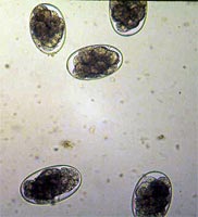
Figure 8. Worm eggs in fecal exam.
The FEC (specifically for H. contortus) has been shown, for the most part, to reflect the animals’ worm burden and also serves as an indicator of seasonal changes in level of infection. Trends in FEC over time can be seen, thus reflecting the relative direction of infection. When worms other than H. contortus predominate, FEC is a less accurate predictor of adult worm burdens.
It is important to know that if heavy infection occurs over a short period of time (1-2 weeks) with Haemonchus, animals may lose substantial amounts of blood with few eggs in the feces as the prepatent period is about 3 weeks.
Blood packed cell volume
Nematode parasites can affect an animals’ ability to maintain erythropoesis (making red blood cells). The PCV is the percent of the blood that is red blood cells and normal is usually above 30%. When PCV drops below 20%, symptoms of anemia usually start to appear. PCV is determined by centrifuging blood in a capillary tube (similar in size to a ball point pen refill) which packs the cells and percent is measured. All nematode parasites can result in chronic anemia where red blood cells are not being made fast enough to keep up with demand. Of special note, H. contortus can lead to substantial acute blood loss and death. PCV values have been used to support other response criteria, and is not necessarily used as a “stand-by-itself” diagnostic tool.
Anemia and FAMACHA©
Level of anemia can be roughly evaluated by observing the color of mucous membranes which are areas where there are a lot of capillaries (very small blood vessels) close to the surface so that tissue color reflects blood color. Such areas are inside the lower eyelid, the gums (only where pigmentation is not present) and inside the vulva. If such membranes are pale (essentially white), impending death is near and deworming is indicated immediately.
The FAMACHA© eye color chart system (Figure 9) was developed in South Africa to help producers monitor and evaluate level of anemia without having to rely on laboratory testing. In this method, the lower eyelid mucous membranes are examined and compared to a laminated color chart bearing pictures of sheep eyes at 5 different levels of anemia.: 1 (red, non-anemic); 2 (red-pink, non-anemic); 3 (pink, mild-anemic); 4 (pink-white, anemic); 5 (white, severely anemic). Since anemia is the primary pathologic effect from infection with H. contortus, this system can be an effective tool for identifying those animals that require treatment (but only for H. contortus).
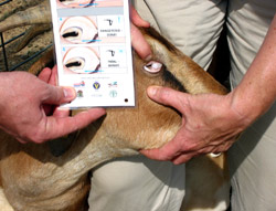
Figure 9. FAMACHA© eye color chart being used to check level of anemia.
FAMACHA© has been extensively tested in South Africa and now the US with excellent results. It has been shown that where animals have been examined at weekly intervals and salvage treatments only were administered, up to 70% of adult animals may not require deworming and only a few required more than one treatment. Compared to previous treatment regimens, total number of treatments may be decreased by up to 90%. Since most of the worms would not be exposed to dewormers, this reduces the development of dewormer resistance. Information on FAMACHA© and training workshops (held in many localities) can be found on the website of the Southern Consortium for Small Ruminant Parasite Control (SCSRPC, www.scsrpc.org).
Worm count and identification
The most absolute and direct method for documenting the number of worms present in an animal is to open it up and collect, identify, and count the worms present. When an animal dies, this can only be done by a properly trained veterinarian or other professional and it might be very expensive. However, one can get an idea of the magnitude of Haemonchus infection by looking for the worms that are visible on the lining of the abomasum. It should be noted that for this to be of any value, the animal can not have been dead for very long. The fresher the animal is after death, the greater the chance to find worms because after death, the worms will move as far down the gut as they can get and eventually die. It is important to note that Telodorsagia and Trichostrongylus are too small to see except under a microscope. Even if thousands of these worms are present, they cannot be seen by the naked eye while mixed in with the gut contents.
Next
Module Home
Certification Table of Contents
Browsing Table of Contents