Gastrointestinal nematodes (worms)
Although there are a number of worms found in goats, only the predominant and usually the most pathogenic ones will be discussed.
General life cycle
Before control measures can be considered, it is important to understand some aspects of the life cycle of these worms. The life cycle consists of part of their life being spent inside the goat and part of their life on the pasture (Figure 1).
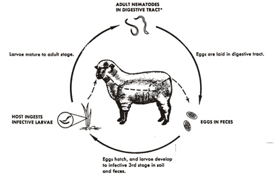
Figure 1. General life cycle of gastrointestinal worm parasites.
Worms mate in the host and females lay eggs that pass out in the feces. The eggs hatch and develop to infective larvae while remaining in the feces. The infective larvae then move out of the feces onto the surrounding forage (Figure 2) where they can be consumed during grazing thus completing the cycle. The time from ingestion of infective larvae to egg laying adults, called the prepatent period, is about three weeks and the time for development from egg to infective larvae can be as short as 7-10 days (especially during the summer months), therefore, transmission (reinfection) and continual pasture contamination can be quite rapid. During the colder months, however, larval development on pasture is delayed and may take up to a month or two to reach the infective larvae stage, thus pasture contamination and reinfection is minimized.
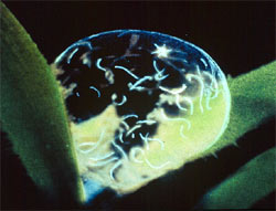
Figure 2. Infective larvae in dew drop on forage.
The infective larvae have a protective sheath making them relatively resistant to adverse environmental conditions and can survive for months, thus extending transmission potential. As long as the temperature and moisture conditions remain warm and wet (especially following periods of substantial rainfall), development and survival continues and pasture contamination accumulates, but if the temperature gets too hot/cold and/or the moisture conditions become dry, development and survival are threatened and pasture contamination dissipates. Transmission of parasites can be reduced by implementing control measures to eliminate the worms from the goat (deworming) and/or reducing the chances that infective larvae have to reinfect the goat (management). Depending on the worm species, the time of the year that is most favorable for transmission varies. This will be addressed below.
Epizootiology
Another way to look at the life cycle is in four phases. Phase 1 is the Parasitic Phase which is the interaction between the goat and the parasite. Phase 2 is the Contamination Phase which is the result of eggs that are passed in the feces during defecation. Phase 3 is the Free-Living Phase when larval stages develop and survive. Phase 4 is the Infection Phase when available infective larvae are consumed during grazing. There are a number of factors that affect what happens and influences control strategies during each of these phases (Figure 3).
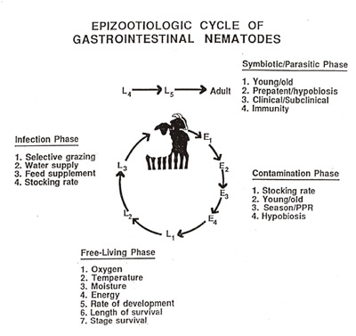
Figure 3. Epizootioligic cycle of gastrointestinal nematodes.
Phase 1- Parasitic Phase
During Phase 1, the parasite has to develop and survive in the host. After ingestion, infective larvae lose their protective sheath and invade the mucosa (lining) of the abomasum, small intestine or large intestine depending on what worm is involved. While in the mucosa, larvae develop to the next larval stage and then return to the surface of the gut mucosa where they become adult worms. The goats major defense mechanism against parasites is the immune system. When infectious agents enter the body, the immune system reacts through a series of activities that mobilize various components (antibodies, killer cells, etc.) that then attack and kill the invaders. These components act on the larval stages in the mucosa and the adults. How strong the immune response is depends on several factors. The immune system has to mature with age, therefore, young animals are relatively susceptible to infection and become more resistant with age. So, young animals usually harbor the heaviest infection levels and suffer the most severe consequences. Adult animals have developed stronger immunity and harbor lower infection levels. One way infection level is measured is by quantifying the number of eggs being passed in the feces. So, relatively high and low egg counts are usually seen in young and adult animals, respectively. Young animals are more subject to clinical disease where signs of infection (diarrhea, rough hair coat, anemia, weigh loss, bottle jaw, etc.) are seen. In older animals, infection usually becomes more subclinical where the only subtle sign may be reduced weight gain. However, nutrition (as mentioned above) and/or stress can alter a host’s immune competence. Under poor nutrition and/or stressful conditions, the immune system loses some effectiveness and can not respond adequately. Therefore, no matter what the age of the animal, the effects of infection will become worse. The prepatent period of most worms is about 3 weeks, but this period can be extended for worms that have the capability to enter a period of delayed or arrested development called hypobiosis. This occurs during the season of the year when the environmental conditions are unfavorable for development and survival of the free-living larval stages. In warm climates, this happens either during summer or winter depending on the worm. In colder climates, all worms capable of hypobiosis will arrest in the winter.
Phase 2- Contamination Phase
The magnitude of pasture contamination during Phase 2 is affected mainly by stocking rate (number of animals per grazing area), age of the animals, season of the year and hypobiosis. The higher/lower the stocking rate, the more/less feces are deposited on the grazing area, thus more/fewer eggs. More eggs are also passed from young vs. older animals. Most worms have a definite seasonality, so during their ‘season,’ more eggs are produced and passed. Of particular note in small ruminants, is a phenomena called the peri-parturient rise (PPR) in fecal egg output. This occurs at or around parturition (kidding) and extends through most of the lactation period. Because parturition and lactation are stressful conditions, the dam’s immune system is compromised. Furthermore, nutrients are partitioned preferentially to support mammary and fetal development and then lactation, which also decreases the animals’ ability to generate an effective immune response to worm infection. This allows the existing female worms to increase the number of eggs laid, thus increasing the number of eggs deposited in the feces. If a worm species undergoes hypobiosis, the development time to the adult stage is extended by several months. This will result in fewer adult worms over time and fewer eggs deposited in feces. However, when these hypobiotic larvae resume development, massive numbers become mature adults over a short period of time and the resultant egg production and deposition in the feces can be very high as well as having severe adverse effects on the animal.
Phase 3 – Free-Living Phase
Development and survival of the free-living stages during Phase 3 depends on prevailing environmental (temperature and moisture) and nutritional (oxygen and energy) conditions. Initially, the first stage larvae develops in the egg which then hatches, and then development and survival to second-stage and finally third-stage (infective) larvae occurs within the fecal mass. The first- and second-stage larvae are unprotected and need oxygen and energy (feed on nutrients and microorganisms) to grow. The infective larvae is enclosed in a protective sheath and does not feed. Temperatures conducive for normal development and survival are between 65-85EF. The lower or higher the temperature gets, development and survival is reduced. Moisture is also crucial for development and survival. Because the initial development and survival occurs within feces, moisture is usually adequate to complete development to the infective larvae; however, if the feces dries out quickly, due to high temperatures and/or physical disruption, the first- and second-stage larvae are susceptible to dessication and will die. If feces remain intact, retain some moisture and do not get too hot or too cold, infective larvae may remain alive for months. A moisture medium (rain/dew) is necessary for infective larvae to migrate out of feces, and they are relatively resistant to environmental conditions encountered due to their protective sheath. Temperature is usually the only factor that may adversely affect the infective larvae. Generally, infective larvae can survive very low temperatures, but may die off during hard freezes. Sustained temperatures above 95EF are usually lethal. The moisture conditions at ground level under forage cover usually is adequate for infective larvae to move around and survive. Since they don’t feed, their length of survival depends on how fast they use up their energy reserves. So, the hotter it is, the faster they move and use up energy stores and survival is shorter. Eventually, infective larvae move up and down the forage when there is a moisture medium (i.e. advancing and receding dew). Rain also provides a moisture medium for larval movement on forage. For the most part, infective larvae do not move much past 12-24 in from feces or 2-3 in up the forage. So, the lower the animals graze and the closer to feces, consumption of infective larvae is increased and vice versa.
Phase 4 – Infection Phase
Phase 4 is affected again by stocking rate in 2 ways. If the same animals are grazing, the stocking rate determines how many eggs initially contaminated (Phase 2) the pasture and, consequently, how many infective larvae will be available for consumption. If the initial contaminating animals are removed and replaced by new animals, the new stocking rate will determine the level of exposure each animal has to infective larvae during grazing, i.e. the higher the stocking rate, the more chance of exposure and vice versa. It is well known that grazing animals usually do not graze close to feces so the further the distance between fecal deposits, exposure is reduced. Eventually feces disintegrate, forage grows well with the fertilization and animals will graze over the area where exposure can be high. Natural sources of water, such as streams, ponds or lakes provide moisture along the banks where forage can grow readily. When animals congregate to drink and consume the attractive forage, defecation in these areas usually leads to increased contamination and eventually more infective larvae. The same can be said for areas where supplements, especially hay, are fed on the ground if conditions are right for development and survival of the free-living stages. Similarly, trees provide an area for animal congregation and shade. Under all these situations, essentially a high stocking rate has been artificially created in a relatively small area where forage is kept closely grazed.
Abomasal worms
Haemonchus contortus (Barberpole worm)
Haemonchus contortus is a voracious blood feeding worm (Figure 4). It gets its name due to the barberpole appearance consisting of the white ovaries that twist around the red blood filled gut (Figure 5). This worm is rather large compared to other stomach and intestinal worms of goats, measuring up to 3/4 of an inch. When large numbers are present, worms can readily be seen as thin (diameter of a paper clip wire) red hair-like worms on the stomach surface (Figure 6). Female worms are prolific egg laying machines and in large numbers with favorable conditions, they can contaminate the environment with a very large number of eggs. These worms thrive under hot and moist environmental conditions, which are conducive for survival and development of the free-living stages, and are found predominantly in tropical and subtropical regions of the world. In the US, these conditions prevail in the southeast. However, in the rest of the US where similar environmental conditions are encountered during the summer, H. contortus transmission also frequently occurs.
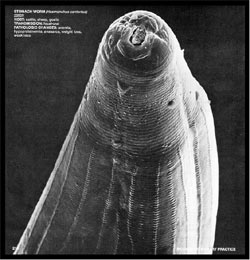
Figure 4. Head of Haemonchus contortus showing lancet that is used to initiate blood flow for feeding.
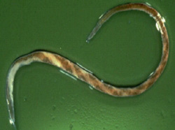
Figure 5. Haemonchus contortus showing barber-pole appearance with white ovaries twisted around red, blood-filled gut.
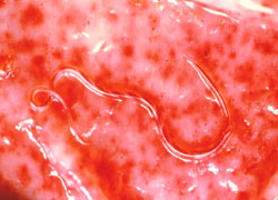
Figure 6. Haemonchus contortus on stomach surface showing areas of hemorrhage.
Generally speaking, H. contortus transmission and infection is at the lowest level during the winter. Transmission and infection increases with the warmer temperatures and increasing moisture during the spring and peaks during the summer. As temperatures and moisture dissipate during the fall, transmission and infection decreases. Hypobiosis has not been observed to occur to any great extent in the SE US because the life cycle can be maintained year around, but it does occur in more northern/western temperate (cold/dry) regions of the US.
Animals infected with H. contortus show symptoms associated with blood loss (anemia), which include pale mucous membranes (most visible by viewing inside the lower eyelid) and bottle jaw (an accumulation of fluid under the chin) (Figure 7). The greater the infection level the more blood is lost and eventually the animal may die.
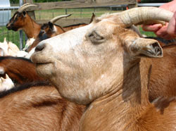
Figure 7. Bottle jaw – accumulation of fluid under the chin.
Telodorsagia (Ostertagia) circumcincta (Brown stomach worm)
The other abomasal worm of importance is Telodorsagia circumcincta which is smaller than H. contortus and is not readily visible since it is about as big as an eyelash. These worms feed mostly on nutrients in mucous and do not feed on blood, per se, but can ingest some blood if present. Female worms do not produce as many eggs as H. contortus. Infection causes direct damage to the stomach lining thereby interfering with digestion and appetite. Infection is usually considered a production disease as animals do not grow very well. However, under very high infection conditions, death can result. When infections reach levels that cause disease to be seen, the primary symptom is diarrhea. This worm thrives in cooler wet environmental conditions which are encountered in the more temperate regions of the US (excludes most of the SE). Hypobiosis occurs when environmental conditions are too cold (winter) or too dry (summer).
Small intestinal worms
Trichostrongylus colubriformis (Bankrupt worm)
Trichostrongylus colubriformis is a very small threadlike worm and is the most predominant small intestinal worm. It is found in goats throughout the US, but seems to thrive better under more cool and wet conditions similar to T. circumcincta. However, in the southeast US, this worm is the next most common and important after Haemonchus and on some farms can cause considerable problems. As with Telodorsagia, this worm feeds on nutrients in mucous and interferes with digestive function resulting in diarrhea. It is called the bankrupt worm because death is seldom the end result and animals just become poor doers leading to loss of production and income.
Nematodirus spp. (Long-necked bankrupt worm)
Nematodirus spp. are relatively large worms (easily seen) and can be found in goats throughout the US although usually in rather small numbers. Problems are rare in the southeast, but in cooler areas of the US there is a possibility of greater numbers of worms accumulating. If heavy infection occurs, production and income losses will result (similar to that of T. colubriformis).
Large intestinal worms
Oesophagostomum spp. (Nodular worm)
Oesophagostomum spp. are relatively large (easily seen) worms and can be found in goats throughout the US, usually in rather small numbers. These worms feed on blood and can contribute to the overall anemia being caused by H. contortus. Although this worm resides in the large intestine, the larvae are found in the mucosa of both the small and large intestine where they form nodules, thus the name nodular worm. Once the larvae leave these nodules they reside in the large intestine.
Trichuris spp. (Whipworm)
Trichuris spp. are usually found in small numbers and the posterior end of the worm is rather large and can be seen. The anterior end of the worm is thread-like, thus the name whipworm. These worms are also blood feeders and, like Oesophagostomum, contribute to the overall blood loss due to other worms. Female worms produce characteristic ‘football’ shaped eggs with protruding plugs at each end.
Next
Module Home
Certification Table of Contents
Browsing Table of Contents