Once any method of assisted animal reproduction is administered, the producer will want to make some attempt at pregnancy confirmation. There are a number of means in which this can be accomplished.
Bumping
Bumping is a method practiced by some producers with a marginal amount of success. The theory behind the technique is to attempt to detect the fetus by way of its “firmness” within the abdomen of the doe. The likelihood of success using this method largely depends on the stage of pregnancy of the doe when the technique is performed, the number of fetus(es) present, and what position the fetus is in at the time the attempt. This method is one of the least reliable for an accurate determination of pregnancy.
Vulva examination
Vulva examination is a method widely used by producers with long-term experience working with both pregnant and open (not pregnant) does. Although not easily recognized until late in the doe’s pregnancy, the skin in and around the vulva becomes more placid, stretchy, and loose in late gestation. When compared to does of the same general age and size that are not pregnant, it can be very apparent that the vulvar region of the pregnant doe is undergoing changes that will allow for successful delivery of the kid. These vulvar changes are usually coupled with noticeable signs of mammary development.
Although certainly not used with any proven reliability until late in gestation, this method is widely practiced and, when asked, experienced producers seem to be of the opinion that the method does work more often than not.
Cervical examination
Producers make some claim that when the cervix of a pregnant doe is examined, a “gray plug” can be seen. This observation has reportedly been made as early as 30 days following conception. An explanation of such an observation may be attributed to the following. The cervix is the “gateway” to the uterus and during pregnancy it has the job of blocking entrance to the uterine body. The claim of a “gray plug” is supported by research proving that during pregnancy while under the influence of progesterone the mucus of the pregnant female becomes quite viscous and thick. The mucus can become so thick that during gestation it acts like “glue” holding the folds of the cervix together so that foreign material cannot enter the uterus. This barrier is commonly referred to as the “cervical seal of pregnancy.” Any disruption of this barrier or seal can, and often will, result in abortion. The cause of the abortion is directly related to microorganisms gaining entry to the uterus and causing infection that leads to subsequent embryonic death.
Ultrasonography
For the diagnosis of pregnancy in goats there are three commonly used ultrasound techniques, each with its own specifically designed piece of equipment. The first and most reliable method is referred to as B-Mode, followed by A-Mode, and finally the Doppler machine. All three methods require that the doe be shaved high on the right side of the abdomen just in front of the mammary gland to remove all hair that may interfere with data received by the ultrasound probe. A coupling gel is also used to ensure a clear projection of information to and from the probe.
If the equipment is not owned and operated by the producer, the cost of ultrasound can vary to a large degree depending on the practitioner, distance traveled, and number of animals to be examined. All three ultrasound methods are highly dependent on the experience and technical knowledge of the operator/technician for a correct diagnosis of pregnancy, i.e., openness (not pregnant), or pseudo pregnancy with hydrometra or muco metra (false pregnancy, e.g., uterine development lacking a fetus).
The most popular method and that offering the most accuracy is the use of B-mode or real-time transabdominal ultrasonography. This method is commonly performed in some states by licensed technicians and often by licensed veterinarians. Most practitioners find that detection is easiest made when the doe is between 45 and 90 days of gestation. Some well-experienced practitioners can determine identification of a fetus as early as 27-30 days, and if examined slightly later, the number of fetuses in the uterine body. Pregnancy can still be identified by means of the caruncles very late in gestation, although actual evidence of the fetus itself may be disrupted by the obstacles in and around the abdominal region of the doe. It is not unusual to see a fetus move about due to the stimulus created by the ultrasonic sound waves. Producers should be aware that there is some minimal risk of fetal abortion if an inexperienced practitioner uses an annular array (transrectal) probe for this purpose. External sector scanners or linear array (transabdominal) probes offer little to no risk of abortion and are preferred by most technicians.
![]()
Transabdominal ultrasonography
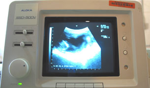
Ultrasound image
A-Mode ultrasound, although not nearly as accurate as the linear interpretations used in B-Mode ultrasonography, is an inexpensive means of pregnancy diagnosis with greatest accuracy achieved at 30-40 days gestation. The A-Mode ultrasound is designed to detect a large body of fluid within the doe’s abdominal cavity. This method can often produce a misdiagnosis by mistaking a full bladder or fluid filled uterus for a pregnant uterus.
The Doppler ultrasound is also used. This machine is designed to detect blood flow by way of a fetal heartbeat. Although cost effective, its degree of accuracy is sometimes less than desirable.
A list of some companies offering ultrasound equipment is listed at the end of this module.
Blood sampling/assay
Although not the least expensive (about $10-15.00 per sample), blood sampling for hormone or protein assay offers by far the greatest degree of safety to the fetus and is one of the most reliable pregnancy detection methods. Results can most often be expected within 5-7 days of the sample’s receipt by the laboratory.
The protocol is very simple, a blood draw of at least 5 cc is made into a sterile, gray or red-topped blood tube as early as 26 days after breeding. Once the laboratory receives the sample, it is screened for a protein in the blood that is only produced by the placenta. This protein, Pregnancy-Specific Protein B (PSPB), can only be present if the doe has, or has very recently, been supporting a viable placenta.
Producers should be aware that a possible false positive could be generated in the event of recent embryonic death enabling residual PSPB to still be detected. False negatives occur most commonly when samples are drawn too early for PSPB detection. Enough time must be given, once the doe becomes pregnant, for the body to build sufficient levels of PSPB in the blood for laboratory detection (26 days from the breeding date).
The screening for fetal protein (PSPB) is a highly reliable test and is most probably only compromised by poor timing in the collection or handling of samples and their cross-contamination. The producer should take care in the harvesting of the whole blood so that no mislabeling or cross-contamination between animals occurs.
A test of non-pregnancy through progesterone analysis can also be made. The corpus luteum of the goat produces measurable levels of progesterone throughout gestation. Milk, serum, and plasma may be analyzed for progesterone concentration. If none is detected, the presence of a functional corpus luteum is unlikely thereby diagnosing the doe as not pregnant. Blood serum and plasma are superior to milk for progesterone analysis. It has been reported that commercial on-farm cattle progesterone test kits have been used in goats with good accuracy.
A list of companies offering laboratory detection of PSPB can be found at the end of this module.
Next
Module Home
Certification Table of Contents
Browsing Table of Contents